
Size Selection of Cells and Microcarrier Beads
CELLNETTA Application Case Studies
In cell-based experiments and cell processing, various operations are used to remove
foreign bodies from cell suspensions. These operations include preprocessing for cell analysis using
a cell sorter and the fractionation of cells and beads after microcarrier cell culture.
Here, we will introduce the method used at Murata Manufacturing to remove beads from HL-60 cell
suspensions.
Implementation method
- (1)Prepare a suspension of HL-60 cells and beads.*1
- (2)Place 40-µm mesh CELLNETTA into a centrifuge tube (Figure 1, Operation 1).
- (3)Apply hydrophilic treatment to the CELLNETTA.*2
- (4)Pour the suspension through CELLNETTA (Figure 1, Operation 2).
*1 Conditions for suspension preparation
| Cell | Type | HL-60 |
|---|---|---|
| Size | 14 µm | |
| Cell count | 5.64×10⁷ cells | |
| Cell concentration | 1.41×10⁶ cells/mL | |
| Bead | Type | Cytodex 3(Cytiva) |
| Size | 120-180 µm | |
| Particle count | 7200 beads | |
| Solvent | Type | RPMI medium |
| Volume | 40 mL |
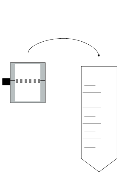
Place into a centrifuge tube
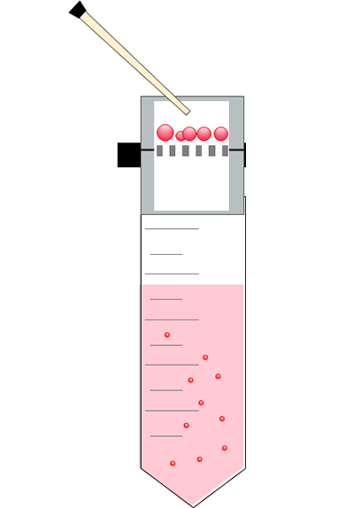
Pour the suspension
*2 For more information, please refer to the “Hydrophilic Treatment Manual“ in the CELLNETTA User Guide.
Results
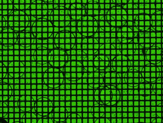
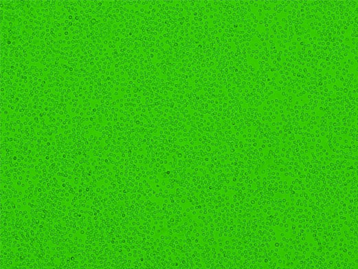
An image of the suspension after processing using CELLNETTA is shown in Figure 2.
After processing using CELLNETTA, the beads in the cell suspension are trapped in CELLNETTA
(Figure 2-A).
Then, by filtering the suspension through the CELLNETTA, only the cells are fractionated (Figure 2-B).
In addition, results of cell measurement of the suspension passed through CELLNETTA showed that
HL-60 cells passed through with high efficiency (95 %) (Table
1).
These results indicate that using CELLNETTA can fractionate beads from cell suspensions.
This simple bead removal method using CELLNETTA is expected to be useful for the fractionation process of cells and beads after bead culture.
| Input solution | Suspension after processing using CELLNETTA | ||
|---|---|---|---|
| Cell | Cell count | 5.64×10⁷ cells | 5.35×10⁷ cells |
| Cell concentration | 1.41×10⁶ cells/mL | 1.35×10⁶ cells/mL | |
| Bead | Particle count | 7200 beads | None observed |
| Solvent | Volume | 40 mL | 39.5 mL |
Product used in this application note
| Pore size | Gamma Irradiation | Product number (P/N) |
|---|---|---|
| 40 µm (custom made) | Gamma Irradiated | MZM1B040B50G |
Notes
- ●This product is not a medical device.
- ●This product is a sample for evaluation purpose.
- ●Please do not ship out your completed product with the sample.
- ●We shall not be liable for any claims on the sample in case it is shipped out to the market.
CELLNETTA Details
For purchasing inquiries, click here. For product inquiries, click here.
This product is single-use only.
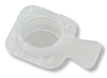
- Compatible with 50 mL centrifuge tubes
- Individually packaged
- Made in Japan
- Gamma irradiated
| Size compatible with 50 mL centrifuge tubes | |||
|---|---|---|---|
| Pore Size (μm) | Package Quantity | Part Number | Application Examples |
| 5 | 1 | MZM1B005B50GA | Purification of culture medium from cell suspensions Removal of red blood cells from whole blood/PBMC |
| 5 | MZM1B005B50GB | ||
| 10 | 1 | MZM1B010B50GA | Purification of single cells |
| 5 | MZM1B010B50GB | ||
| 15 | 1 | MZM1B015B50GA | Fractionation of clusters and single cells |
| 5 | MZM1B015B50GB | ||
| 20 | 1 | MZM1B020B50GA | Fractionation of clusters and single cells |
| 5 | MZM1B020B50GB | ||
| 100 | 1 | MZM1B100B75GA | Spheroid size-based sorting for drug efficacy testing |
| 5 | MZM1B100B75GB | ||
| 200 | 1 | MZM1B200B75GA | Fractionation of microcarrier beads Spheroid size control |
| 5 | MZM1B200B75GB | ||
*We recommend selecting a pore size slightly smaller than the target cells. Please contact us
for guidance on selecting the optimal pore size for your application.
*Flow behavior may vary depending on cell morphology, flow conditions, and other
factors.