
Size selection of cell clusters and single cells~Application to CTC research~
CELLNETTA Application Case Studies
There are two types of circulating tumor cells (CTCs) that circulate in the bloodstream: single cells and multicellular groupings called clusters. There has been more focus on the difference in metastatic potential of cancer between these two types. Here, we will introduce a case study of using CELLNETTA for the fractionation of simulated single-cell CTCs and cluster CTCs prepared using floating culture with fluid shear stress, conducted by Dr. Manabu Maeshiroand Associate Professor Satoru Shinrikiof the Department of Molecular Laboratory Medicine at the Faculty of Life Sciences of Kumamoto University (As of October 2019).
*The component cells of clusters fractionated using CELLNETTA have been shown to efficiently form CTC clusters in mouse blood (Maeshiro M Shinriki S Liu, R et al Colonization of distant organs by tumor cells generating circulating homotypic clusters adaptive to fluid shear stress Sci Rep 11 6150 2021 doi 10 1038 /s 41598 021 85743 z)
Implementation method
[CELLNETTA processing method]
- (1)Culture a human oral squamous cell carcinoma cell line (SAS) of 3.0×10⁶ cells in 15 mL of medium (15 mL centrifuge tube) in a rotary suspension culture.
- (2)Apply hydrophilic treatment to the CELLNETTA. *1
- (3)Process the cell suspension in portions not exceeding 2.0 x 10⁵ cells using a 20 µm mesh CELLNETTA.
- (4)Recover the cells trapped by CELLNETTA through backwashing.
- (5)Repeat steps (2) to (3) five times to process the entire suspension.
- (6)Recover and combine all the flow-through, and further process it using a 15 µm mesh CELLNETTA in the same way as in steps (2) to (4), dividing it into five portions.
[Method of analysis]
Photograph the mesh section of CELLNETTA under a microscope (×100) and measure the number of
cell clusters and single cells trapped by the mesh.
Evaluate five visual fields for each of A, B, and C (Figure
1), and calculate the average.
Analyze the flow-through in the same way by evaluating three visual fields for the 20 µmmesh
and one visual field for the 15 µm mesh.
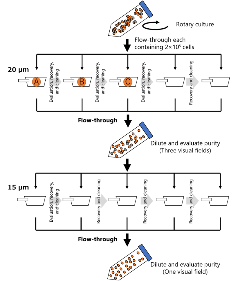
*1 For more information, please refer to the “Hydrophilic Treatment Manual“ in the CELLNETTA User Guide.
Results
The abundance of cell clusters trapped using the 20 µm mesh was approximately 78 % (Figure 2A).
However, flow-through also contained approximately 34 % clusters, the majority of which were
doublets (Figure 2B).
Flow-through was subsequently processed using the 15 µm mesh to ensure the purity of single
cells.
As a result, only approximately 4 % clusters were observed in the flow-through, almost all of
which were single cells (Figure 2C).
When the cell clusters trapped using the 20 µm mesh were recultured, no issues in cell adhesion
or cell growth were found (Figure 3).
These results suggest that cells trapped using the 20 µm mesh and the cells trapped from flow-through using the 15 µm mesh are suitable for analysis as cluster cells and single cells, respectively.
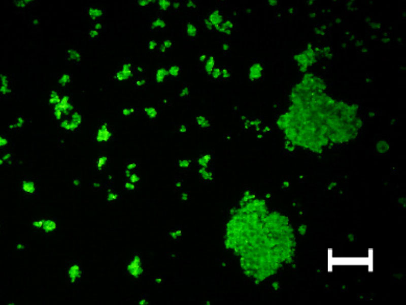
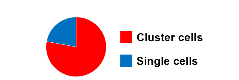
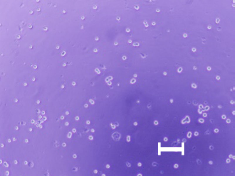
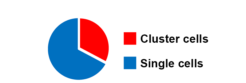
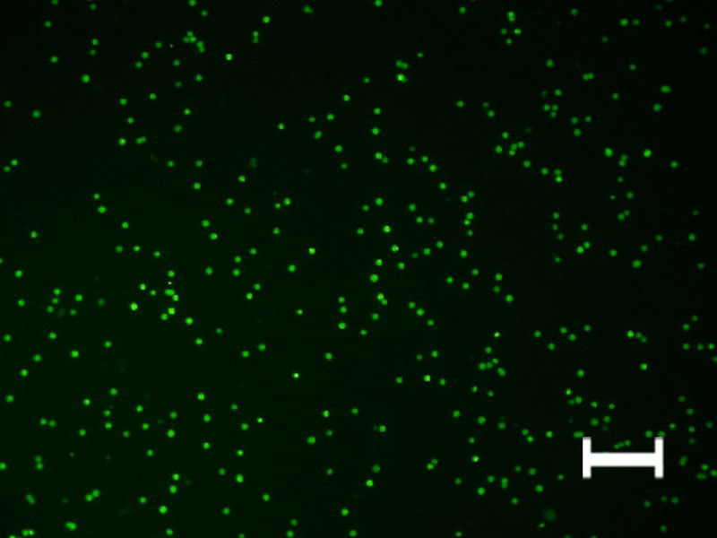
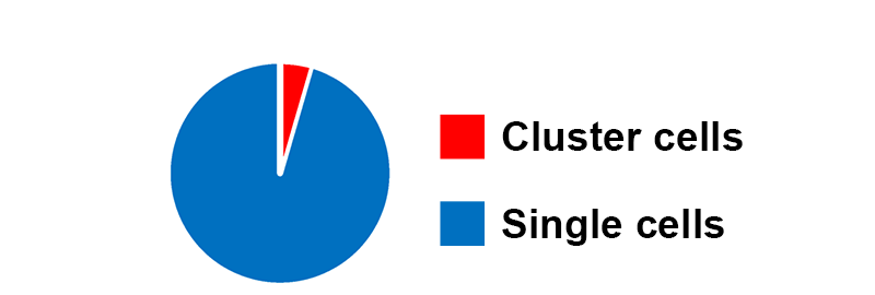
Micrographs of the cells obtained in each operation and the abundance of clusters. After 12 hours of rotary suspension culture, the abundance of cell clusters trapped using the 20 µmmesh was approximately 75 %. The flow-through using the 15 µmmesh consisted almost entirely of single cells, with approximately 4 % cluster cells.
Cluster abundance (%) = 100 ×(clusters)/(single cells + clusters). The scale bar is 100 µm.
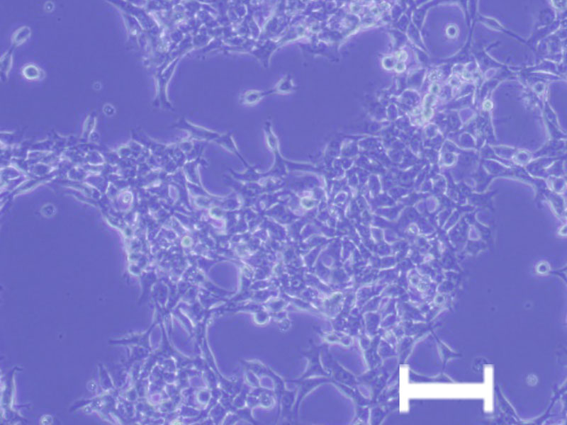
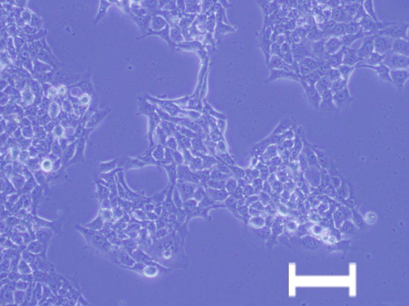
No problems with cell adhesion or cell growth were found. The scale bar is 100 µm.
Product used in this application note
| Pore size | Gamma Irradiation | Package Quantity | Product number (P/N) |
|---|---|---|---|
| 15 µm | Gamma Irradiated | 1 | MZM1B015B50GA |
| 5 | MZM1B015B50GB | ||
| 20 µm | Gamma Irradiated | 1 | MZM1B020B50GA |
| 5 | MZM1B020B50GB |
Notes
- ●This product is not a medical device.
- ●This product is a sample for evaluation purpose.
- ●Please do not ship out your completed product with the sample.
- ●We shall not be liable for any claims on the sample in case it is shipped out to the market.
CELLNETTA Details
For purchasing inquiries, click here. For product inquiries, click here.
This product is single-use only.
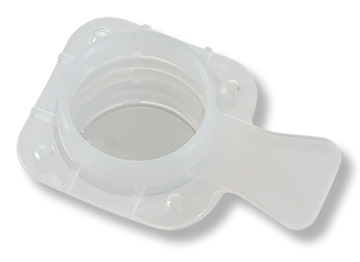
- Compatible with 50 mL centrifuge tubes
- Individually packaged
- Made in Japan
- Gamma irradiated
| Size compatible with 50 mL centrifuge tubes | |||
|---|---|---|---|
| Pore Size (μm) | Package Quantity | Part Number | Application Examples |
| 5 | 1 | MZM1B005B50GA | Purification of culture medium from cell suspensions Removal of red blood cells from whole blood/PBMC |
| 5 | MZM1B005B50GB | ||
| 10 | 1 | MZM1B010B50GA | Purification of single cells |
| 5 | MZM1B010B50GB | ||
| 15 | 1 | MZM1B015B50GA | Fractionation of clusters and single cells |
| 5 | MZM1B015B50GB | ||
| 20 | 1 | MZM1B020B50GA | Fractionation of clusters and single cells |
| 5 | MZM1B020B50GB | ||
| 100 | 1 | MZM1B100B75GA | Spheroid size-based sorting for drug efficacy testing |
| 5 | MZM1B100B75GB | ||
| 200 | 1 | MZM1B200B75GA | Fractionation of microcarrier beads Spheroid size control |
| 5 | MZM1B200B75GB | ||
*We recommend selecting a pore size slightly smaller than the target cells. Please contact us for guidance on selecting the optimal pore size for your application.
*Flow behavior may vary depending on cell morphology, flow conditions, and other factors.