
Removal of debris and red blood cells
for
pre-processing of single-cell analysis
CELLNETTA Application Case Studies
Conventional methods for studying gene expression have been based on analyzing a single
sample of a cell population to obtainaverage data. Single-cell analysis investigates the gene
expression of individual cells.
Single-cell analysis is applied to research on the diversity of cell populations, cell-cell
interactions in diseases, the search for specific marker genes, and the regulatory mechanisms of
drug resistance. The most critical factor in obtaining highly accurate data in single-cell analysis
is the preparation of a high-quality sample that is free of materials (hereafter referred to as
“contaminants”) other than the target cells to be analyzed.
The presence of a large amount of cell debris or red blood cells in the sample will impact data
reliability during single-cell analysis, leading to lost time and money.
The loss of necessary cells during sample preparation is also an issue.
Here, we will introduce a case study of the use of CELLNETTA for the removal of contaminants for
pre-processing of single-cell analysis.
The study was conducted by Dr. Satoshi Ueha, Associate
Professor of the Division of Molecular Regulation of Inflammatory and Immune Diseases at the
Research Institute for Biomedical Science at the Tokyo University of Science. (As of October 2021)
(Also available as a video)
CELLNETTA Application Example Pre-processing for Single-cell Analysis
Implementation method
- (1)Prepare a partially purified cell suspension of mouse lung tissue dispersed by enzymatic digestion. Then, stain it with various cell surface markers and Calcein-AM, a fluorescent dye for staining live cells.
- (2)Apply hydrophilic treatment to the CELLNETTA.*
- (3)Place 5 mL of buffer in each of the three wells of a 6-well plate. Place 3 mL of buffer in one well.
- (4)Place CELLNETTA in wells containing 5 mL of buffer, and add 1 mL of the cell suspension prepared in (1).
- (5)Move CELLNETTA up and down in the buffer 2-3 times, then transfer it to a well containing 5 mL of new buffer. Repeat the process again.
- (6)Submerge CELLNETTA in the well containing 3 mL of buffer and allow cells to float.
- (7)Pipette the suspension approximately 10 times, then recover 1 mL of the suspension.
- (8)Repeat step (7) again to recover a total of 2 mL of cell suspension.
- (9)Stain the cell suspension recovered in step (8) with PI (propidium iodide), a fluorescent dye for staining dead cells, then analyze it with a flow cytometer.
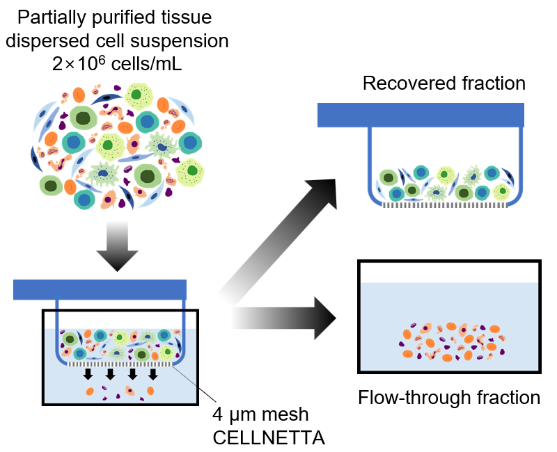
* For more information, please refer to the “Hydrophilic Treatment Manual“ in the CELLNETTA User Guide.
Results
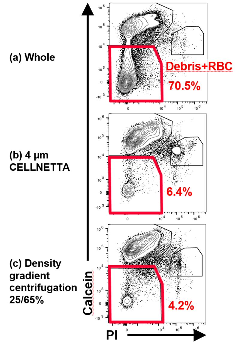
Partially-purified cell suspensions of mouse lung tissue dispersed by enzymatic digestion consist
of 70.5 % contaminants, such as debris and red blood cells (Figure
1(a): Whole). Density gradient centrifugation, which is commonly used to remove such contaminants, reduced the
percentage of contaminants to 4.2 % after processing (Figure
1(b): Density gradient centrifugation 25/65 %). Since there is a difference in size between the contaminants and the target cells, purification
using CELLNETTA reduced contaminants to 6.4 % (Figure 1(c): 4 µm
CELLNETTA).
Moreover, a comparison of the cell recovery rate in suspensions that used CELLNETTA and
centrifugation with respect to cell suspensions prior to processing indicates that CELLNETTA
reduced the loss of each cell type better than density gradient centrifugation (Figure 2).
The processing time of density gradient centrifugation is approximately 40 minutes. However,
with CELLNETTA it was possible to reduce this time to less than 10 minutes.
These results indicate that CELLNETTA can be used as a pre-processing tool for single-cell analysis to quickly and accurately remove debris and red blood cells.
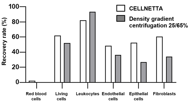
suspension afterprocessing Number of cells in the
suspension priorto processing
Product used in this application note
| Pore size | Multi well plate model | Product number (P/N) |
|---|---|---|
| 4 µm (custom made) | 6 well plate | Please contact us. |
| 12 well plate | Please contact us. |
Notes
- ●This product is not a medical device.
- ●This product is a sample for evaluation purpose.
- ●Please do not ship out your completed product with the sample.
- ●We shall not be liable for any claims on the sample in case it is shipped out to the market.
CELLNETTA Details
For purchasing inquiries, click here. For product inquiries, click here.
This product is single-use only.
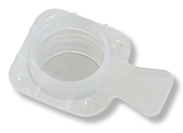
- Compatible with 50 mL centrifuge tubes
- Individually packaged
- Made in Japan
- Gamma irradiated
| Size compatible with 50 mL centrifuge tubes | |||
|---|---|---|---|
| Pore Size (μm) | Package Quantity | Part Number | Application Examples |
| 5 | 1 | MZM1B005B50GA | Purification of culture medium from cell suspensions Removal of red blood cells from whole blood/PBMC |
| 5 | MZM1B005B50GB | ||
| 10 | 1 | MZM1B010B50GA | Purification of single cells |
| 5 | MZM1B010B50GB | ||
| 15 | 1 | MZM1B015B50GA | Fractionation of clusters and single cells |
| 5 | MZM1B015B50GB | ||
| 20 | 1 | MZM1B020B50GA | Fractionation of clusters and single cells |
| 5 | MZM1B020B50GB | ||
| 100 | 1 | MZM1B100B75GA | Spheroid size-based sorting for drug efficacy testing |
| 5 | MZM1B100B75GB | ||
| 200 | 1 | MZM1B200B75GA | Fractionation of microcarrier beads Spheroid size control |
| 5 | MZM1B200B75GB | ||
*We recommend selecting a pore size slightly smaller than the target cells. Please contact us
for guidance on selecting the optimal pore size for your application.
*Flow behavior may vary depending on cell morphology, flow conditions, and other
factors.